Explain the Differences Between Venous and Mixed Venous Blood Samples
Venipuncture samples for venous CO2 content and chloride concentrations were obtained in 336 patients with arterial blood pH PaO2 PaCO2 and oxygen saturation determinations. The normal mixed venous PO 2 PvO 2 is 40 mmHg.

Mixed Venous Oxygen And Carbon Dioxide Content Deranged Physiology
For larger quantities will take venous blood.

. If there is a decrease in cardiac output then that means that the arterial and mixed venous content is different it means that the venous O2 content has decreased low mixed venous content will cause low cardiac output because there are not getting enough O2 to pump it up. Thus the result is an average of venous blood. September 9 2019 Posted by DrSamanthi.
Shunt fraction is the calculated ratio of venous admixture to total cardiac output. Mostly venous blood is drawn in the fasting state. DNA extractions by manual procedure and MP were performed each on cell pellet venous blood and DBS samples and tested by Amplicor HIV-1 DNA assay.
Venous blood was used to prepare cell pellet and DBS samples. In many studies a very good correlation has been shown between venous blood gas and the arterial blood gas. Arterial blood is bright red colour but venous blood is dark maroon in colour.
Scvo2 is more conveniently measured and less risky than Svo2 measurement. This generally produces an SvO 2 of 70. Venous admixture is that amount of mixed venous blood which would have to be added to ideal pulmonary end-capillary blood to explain the observed difference between pulmonary end-capillary PO 2 and arterial PO 2.
In a recent study we reported differences between capillary and venous complete blood counts CBC in healthy term neonates on day 1 of life. Scvo2 measures central venous oxygen saturation level from veins draining the head and upper body while Svo2 measures mixed venous oxygen saturation from the lower half of the body. Arterial blood travels through the left chamber of the heart whereas venous blood moves through the right chambers of the heart.
Significant differences for standard bicarbonate SHCO 3 were found only when jugular and cephalic venous blood were compared with arterial blood in dogs with a metabolic. Since venous blood gas is easy to sample from the peripheral veins or the central veins in patients with central venous catheters it is a more comfortable and an easy procedure for some patients and the physicians. Supporting this hypothesis the difference between Hb measured in capillary and venous blood has been found to be higher in males than females likely mediated by males higher overall body red blood cell count contrasted with the reduced hematocrit red cell count and Hb in their microvasculature system including their capillaries27 43 In the study of over 36000 paired.
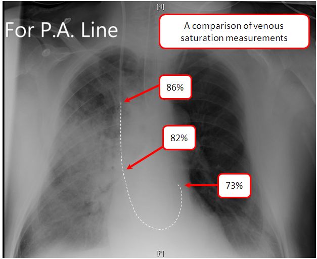
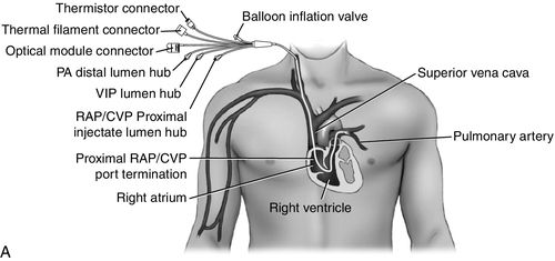
Venous blood venipuncture. If the PvO 2 rises above 60 mmHg the SvO 2 may rise to arterial saturation levels. A mixed venous blood gas is a sample aspirated from the most distal port of the PA catheter offering a mixture of inferior vena cava blood superior vena cava blood and the coronary sinuses.
The median cubital vein is usually preferred. Blood is taken directly from the vein called phlebotomy. The partial pressure of carbon dioxide PCO2 is the measure of carbon dioxide within arterial or venous blood.
It is well known that capillary blood has higher hemoglobin Hb and hematocrit Hct values than venous blood. A forearm site is preferred. The linear correlation between actual calculated arterial HCO3- and the measured venous CO2 was significant P less than 001.
Previous research 48 showed no difference between venous and capillary blood samples using continuous glucose monitoring systems for determining the blood glucose response to. Generally under normal physiologic conditions the value of PCO2 ranges between 35 to 45 mmHg or 47 to 60 kPa. Differences in metabolomes between arterial and venous blood and their associations with gestational length birth weight sex and whether the baby was the first born or not as well as maternal age.
During cardiac arrest tissue hypoxia is all but a certainty and is reflected by the lower pH and higher PCO 2 on the venous side. The blood sample is taken from the forearm wrist or ankle veins. At times venous blood may be obtained using a vascular access device VAD such as a central venous pressure line or an IV start.
The results of this study demonstrated significant differences between the blood from various venous sites and the arterial site for P CO2 and pH in all acidbase states. The arterial blood is bright red in color and the venous blood is blackish red in color. 75 The human body under normal conditions makes far more oxygen available to the cells than the cells use.
The main difference between arterial and venous blood is that arterial blood is oxygenated whereas venous blood is deoxygenated. Venous blood is the specimen of choice for most routine laboratory tests. Aspiration of air into the blood gas syringe during sampling or the presence of an air bubble are potential causes for false elevation of SvO 2.
Venous blood samples from children under 18 months born to HIV-infected mothers between January and April 2008 were collected. The normal capillary and venous hematologic values for neonates have not been defined clearly. In cardiac arrest victims the disparity between arterial and venous values is even greater.
A venous pH of 7 or lower for example predicted an arterial pH of 72 or lower in 98 of cases. Blood is a body fluid that delivers vital substances such as nutrients. The blood is obtained by direct puncture to a vein most often located in the antecubital area of the arm or the back top of the hand.
A sample of venous blood from a healthy normal human would still have an oxygen saturation of about. It often serves as a marker of sufficient alveolar ventilation within the lungs. The key difference between arterial and venous blood gas is that arterial blood gas test uses a small blood sample drawn from an artery while venous blood gas test is a comparatively less painful test that uses a small blood sample drawn from a vein.
Central and mixed venous blood gases offer us a glimpse of whole-body oxygen extraction. Typically the measurement of PCO2 is performed via an arterial.

Comparison Of Minimally And More Invasive Methods Of Determining Mixed Venous Oxygen Saturation Journal Of Cardiothoracic And Vascular Anesthesia

Mixed Venous Oxygen And Carbon Dioxide Content Deranged Physiology

No comments for "Explain the Differences Between Venous and Mixed Venous Blood Samples"
Post a Comment